
Virtual Staining for Fast Histological Imaging
During Cancer Surgery
Product introduction
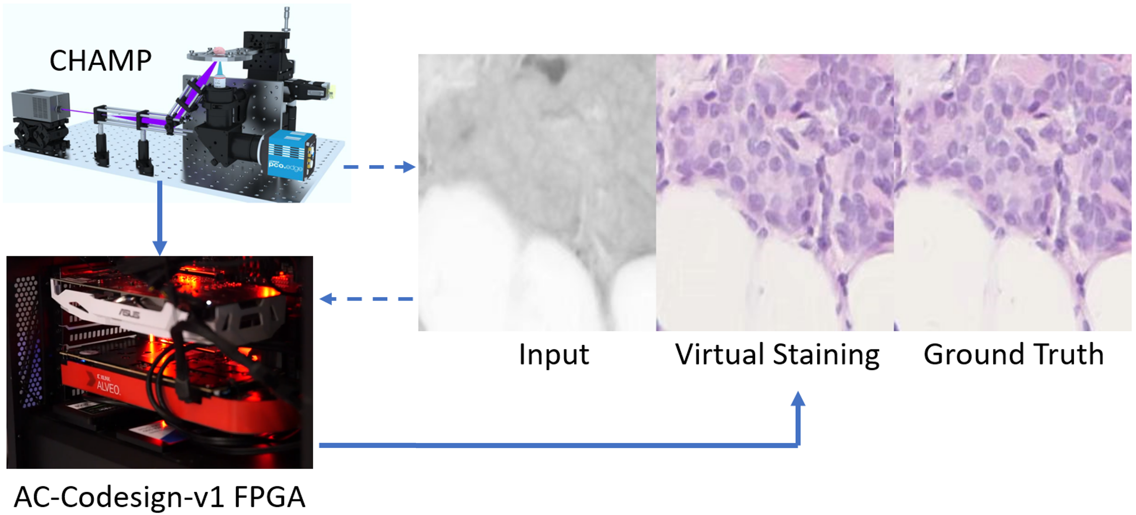
Histological images can reveal rich cellular information of tissue sections, which are widely used by pathologists in cancer surgery. By incorporating ultraviolet photoacoustic microscopy with deep learning, PhoMedics proposed a rapid histological imaging method that can generate virtually stained histological images for fresh tissue specimens.
CHAMP Microscope™ developed by PhoMedics generates high-resolution histological images and uses deep learning algorithms to virtually stain grayscale CHAMP images into accurate histological images.
Technology Specifications
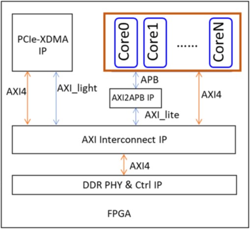
GPU-based systems are unsuitable for compact surgical equipment due to high power consumption. To address this, PhoMedics collaborates with The Center to customize the AI accelerator core of the AC-Codesign-v1. This solution features:
- Algorithm: U-Frame (proposed by PhoMedics)
- Parallel Architecture: 8 AI cores (based on AC-Codesign-v1)
- Peak Compute: 1.6 TOPS (200MHz)
Developed on the Xilinx U250 FPGA platform, this delivers a low-power alternative for surgical environments.
If you are interested in our technologies, please contact us.